There are some things you will need to know when tending to a patient at their bedside. Observing a patient’s features and then diagnosing a potential condition is a key skill of the role. This question bank is designed to test your knowledge of all things observation and bedside related.
Just navigate to the question you want or scroll to take the entire test. Once you have guessed, click reveal to show the answer,
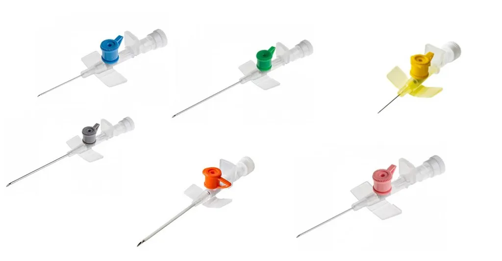
Order the cannula colour with the correct size (G or gauge) from smallest to largest. Remember the lower the gauge, the wider bore the cannula.
So… it goes from smallest to largest: yellow, blue, pink, green, white, grey, orange. I’d be interested to know how this came about. There’s no clear logic to it!
Signs of chronic liver disease
The following are a sign of / associated with chronic liver disease. For each one, choose true or false?
Keiser Fleisher rings: Wilsons disease
Diabetes mellitus and CLD: think Haemochromatosis
SOB and CLD: Think Alpha 1 antitrypsin
Female and CLD and no alcohol: PBC and PSC
Gottrons papules are the papules on the backs of the fingers seen in dermatomyositis.
Auer rods are seen on a blood film and are characteristic of AML.
A McMurray test is the testing to look for damaged menisci in the knee examination.
Endocrine observation and diagnosis
For each following observation, what would be your potential diagnosis?
Random blood sugar 21, urine: ketones, ABG: pH 7.32
Random blood sugar 21, urine: no ketones, ABG: pH 7.42
Diabetes mellitus, poor long term glycaemic control: HBA1c reflects glycosylated haemoglobin and thus DM control over the last few months
MCV 102 (80-99), CK 240 (<150)
Hypothyroidism: watch for the high CK due to abnormalities in the muscle cell membrane. Macrocytosis is commonly caused by Hypo/hyperthroidism, B12 deficiency and alcoholism
Clinic Blood glucose 5.2 mmol/l, HBA1c 6.2%
Diabetes mellitus, good long term glycaemic control: HBA1c reflects glycosylated haemaglobin and thus shows good DM control over the last few months
Lid Lag, Lid retraction, goitre
Graves Disease
Hypothyroidism, no goitre
Amiodarone induced hypothyroidism
Hypertension, striae, moon face
Cushings disease/ syndrome: Remember this fact…
Cushing’s syndrome is the syndrome encompassing all the different clinical entities
Cushing’s disease is the specific release of ACTH from the pituatory itself, thus Cushings disease is one form of the Cushing’s syndrome.
Other causes of Cushings syndrome include: adrenal adenoma
Adrenal carcinoma
Glucocorticoid treatment (e.g. prednisolone in rheumatoid arthritis)
Extreme episodic palpitations with hypertension
Phaeochromocytoma: Tumour that produces catecholamines leading to hypertension, stroke, palpitations and cardiac dysrythmias. Treatment is excision. Diagnosis is based on finding elevated levels of urinary catecholamines followed by imaging of the adrenal glands themselves.
Excessive thirst, normal blood sugar and elecrolytes
Diabetes insipidus: caused by impaired secretion of Anti diuretic hormone (ADH) leading to impaired water retention.
ADH acts exactly as you would expect it to so if you don’t have enough “anti diuretic” the result is diuresis!
Blood tests can be normal. Remember poorly concentrated urine in water depravation can lead to a diagnosis (water depravation test)
Large tongue, prominent supra orbital ridge, changing facial appearance
Acromegaly: Pituatory excessive secretion of growth hormone. Biochemical diagnoses remember IGF1 (insulin like growth factor 1). Gold standard diagnosis is to perform an oral glucose tolerance test and then measure levels of growth hormone.
Paroxsysmal flushing of the face and weight loss
Carcinoid syndrome: excessive release of 5HIAA by this tumour causes paroxsymal flushing and sweats. Diagnosis: 24 hour urine collection for 5HIAA (5 hydroxyindolacetic acid)
Gynecomastia
Marijuana use: other causes include chronic liver disease (alcoholism), testosterone replacement (bodybuilders using drug to combat impotence), other drugs (classically spirinolactone and digoxin)
Pigmented palmar creases, hypothermia
Addison’s disease: Steroid lack secondary to adrenal gland failure of any cause (autoimmune/ cancer infiltration/ TB etc)
Goitre with hypothyroidism
Hashimoto’s thyroiditis: autoimmune infiltration and destruction of the thyroid gland
JVP cannon waves
A 62 year old man has ischaemic heart disease. On inspection of the JVP cannon waves are seen. This is because of
Atrial Fibrillation
Congestive cardiac failure
Ventricular Tachycardia
Asystole
Complete heart block
Complete heart block
In CHB you get asychronous contraction of the atria and ventricles. Common causes include conduction system disease (IHD) and drugs (e.g. combinations of beta blockers and calcium channel blockers).
Deep breaths
A 22 year old female is sat in an A&E triage cubicle talking to her partner. You observe
She has a respiratory rate of 28 She is taking deep breaths in She does not appear to be wheezy She does not look well
The A&E cardex suggests she started a new medicine this morning.
What is being observed?
Kussmaul sign
Traub’s sign
Adverse drug reaction
Anaphylaxis
Respiratory acidosis
Acute asthma
Kussmaul sign
“Kussmaul breathing” – respiratory compensation for a metabolic acidosis.
Traub’s sign is pistol shot femorals on auscultaion of the femorals in sever AR: the nosie as the blood refluxes back through the femoral artery!
Deep breaths 2
Continued from previous question: [A 22 year old female is sat in an A&E triage cubicle talking to her partner. You observe
She has a respiratory rate of 28 She is taking deep breaths in She does not appear to be wheezy She does not look well
The A&E cardex suggests she started a new medicine this morning.
What is her most likely underlying diagnosis from the following list
Aspirin Overdose
Methanol ingestion
Alcohol Poisoning
Traub’s sign
Type I diabetes
None of the above
Type I diabetes
Aspirin OD – yes this would do it
Methanol poisoning – yes this would also do it
Lactic acidosis from pancreatitis – yes this would do it
but…. they’re all much rarer than type I diabetes mellitus presenting in this way (diabetic ketoacidosis).
Respiratory matching condition
Match the following features with their underlying condition.